
Think about the picture quality of the old standard-definition television that you owned maybe 10 or 15 years ago. Now, compare that with today’s high-definition televisions, which allow you to see the texture of an actor’s skin or individual blades of grass on a baseball field.
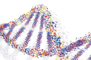
That comparison is roughly equivalent to the progress we’ve seen in diagnostic imaging of the breast in recent years. With advancements such as high-resolution ultrasound and digital tomosynthesis (commonly called 3-D mammography), we are able to obtain far better images of the breast tissue than ever before.
As a result, these imaging technologies are making a major difference in the diagnosis and treatment of breast cancer, which will impact an estimated one in eight women during her lifetime, according to the American Cancer Society. Highly detailed images are allowing us to find smaller and smaller breast cancers, including cancers considered:
- Stage 1 — a tumor 2 cm (about three-quarters of an inch) or less across that has not spread to the lymph nodes or other distant sites.
- Stage 0 —a cancerous, non-invasive condition of the breast called ductal carcinoma in situ (DCIS) in which cancer cells have not yet moved from the milk ducts into surrounding breast tissue.
Detecting cancer at these stages is critical because the 5-year relative survival rate for women with stage 0 or stage 1 breast cancer is close to 100 percent.
Using all of our imaging tools
I can recall one patient, a women in her early 40s, whose case shows the life-saving potential of our new imaging technologies. This patient had been choosing to have annual breast screening exams for several years with traditional digital mammography. Those exams were normal.
One year, she decided to choose digital tomosynthesis or 3-D mammography. It allows us to view the breast from many more angles and actually see in between dense tissues in ways we couldn’t with traditional 2-D images.
This woman’s 3-D mammogram showed an abnormality, so we followed it up with a high-resolution ultrasound exam of her breast. Ultrasound creates images using sound waves. Recent improvements in ultrasound technology provide remarkable detail of structures inside of the body. In this patient’s case, we found a breast tumor that was less than 1 cm.
Her breast tumor was removed during lumpectomy surgery, and her cancer has very likely been cured. Without these new, highly sensitive imaging modalities, it would have been years before her cancer was detected. At that point, her cancer would have been found at a later stage when it would be more difficult to treat and more likely to have spread to nearby lymph nodes.
Treatment without surgery
Breast cancer has traditionally been treated with surgery (lumpectomy or mastectomy) and some combination of radiation therapy, chemotherapy and hormone therapy. Advanced imaging that detects smaller, early stage cancers is now opening up the possibility of treating breast cancer — without surgery.
In fact, CentraState Medical Center is helping pioneer two such treatments that have the potential to give women more options for fighting breast cancer. The hospital is involved in:
- A national clinical trial to evaluate the use of cryoablation without lumpectomy surgery to treat breast cancer. Cryoablation is a nonsurgical procedure that uses image-guidance to locate and freeze a tumor through a tiny incision in the breast. The freezing process may potentially kill the tumor and the dead tissue is removed by the body’s immune system — not unlike the way the body heals a bruise.
- A local study of the effectiveness of using stereotactic needle biopsy to remove small breast tumors. Needle biopsy is commonly used to extract and examine a suspicious mass in the breast to diagnose cancer. The entire mass is then removed during surgery. With our new imaging technologies, we are now sometimes able to remove the entire tumor with the needle, a “stereotactic lumpectomy,” which may eliminate the need for further surgery.
The potential for these treatments is extraordinary. We at CentraState and the Statesir Cancer Center are proud to be on the cutting edge of breast cancer research.
Comprehensive cancer prevention and treatment
The Star and Barry Tobias Women’s Health Center at CentraState offers breast health wellness, diagnostic, treatment and counseling services. In addition to mammography, the center also offers stereotactic needle biopsy, breast needle localization, breast ultrasound and bone densitometry. For more information, click here or call (732) 294-2626.
The Statesir Cancer Center at CentraState delivers comprehensive treatment and support services to help patients and families. Patients also have access to ongoing clinical trials, where appropriate. To learn more, call (855) 411-CANCER.
 Dr. Kenneth Tomkovich is a board-certified interventional radiologist and internationally recognized expert in breast cancer diagnosis and interventional treatment procedures. He is medical director of Breast Imaging and Interventional Radiology at CentraState Medical Center and a physician at Princeton Radiology (formerly Freehold Radiology). Dr. Tomkovich can be reached by calling (732) 294-2946.